Lab Services

A medical laboratory or clinical laboratory is a laboratory where tests are carried out on clinical specimens in order to obtain information about the health of a patient in order to provide diagnosis, treatment, and prevention of disease. Clinical laboratories are thus focused on applied science, mainly on a production-like basis, as opposed to research laboratories that focus on basic science on an academic basis.

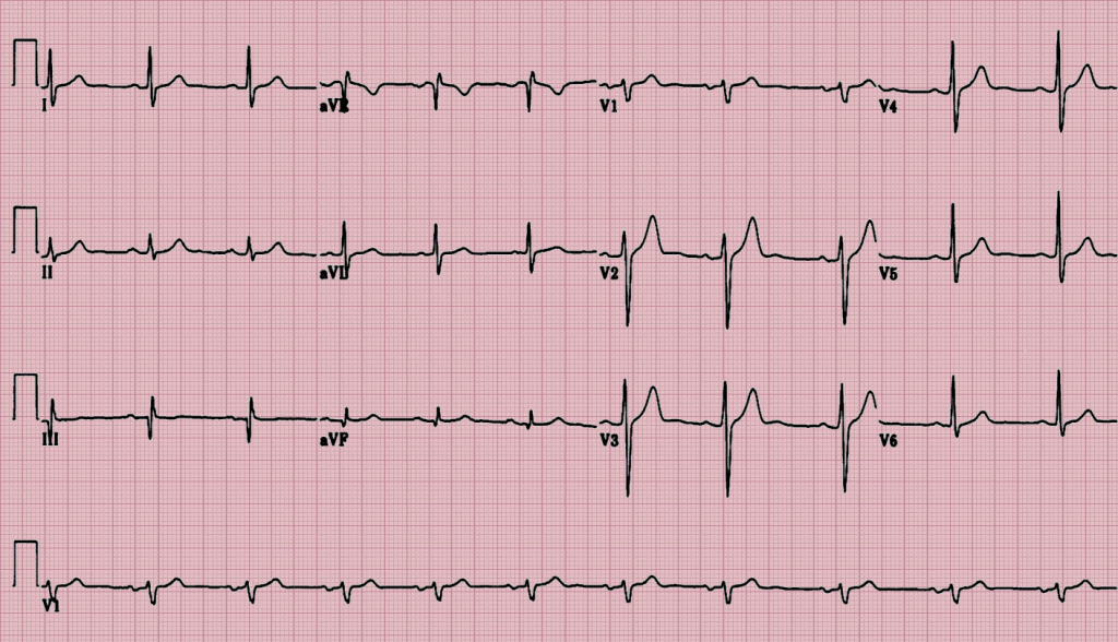
What is an EKG?

Your doctor may suggest you get an electrocardiogram — also called an EKG or ECG — to check for signs of heart disease. It’s a test that records the electrical activity of your ticker through small electrode patches that a technician attaches to the skin of your chest, arms, and legs.

How Should I Prepare?

Some things you can do to get yourself ready:

  • Avoid oily or greasy skin creams and lotions the day of the test because they can keep the electrodes from making contact with your skin.
  • Avoid full-length hosiery, because electrodes need to be placed directly on your legs.
  • Wear a shirt that you can remove easily to place the leads on your chest.

What Happens During an Electrocardiogram?

A technician will attach 10 electrodes with adhesive pads to the skin of your chest, arms, and legs. If you’re a guy, you may need to have your chest hair shaved to allow a better connection.

During the test you’ll lie flat while a computer creates a picture, on graph paper, of the electrical impulses that move through your heart. This is called a “resting” EKG, although the same test may be used to check your heart while you exercise.

It takes about 10 minutes to attach the electrodes and complete the test, but the actual recording takes only a few seconds.

Your doctor will keep your EKG patterns on file so that he can compare them to tests you get in the future.

What Is a CT Scan?

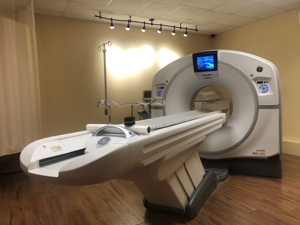
A computed tomography (CT or CAT) scan allows doctors to see inside your body. It uses a combination of X-rays and a computer to create pictures of your organs, bones, and other tissues. It shows more detail than a regular X-ray.

How Do CT Scans Work?

They use a narrow X-ray beam that circles around one part of your body. This provides a series of images from many different angles. A computer uses this information to create a cross-sectional picture. Like one piece in a loaf of bread, this two-dimensional (2D) scan shows a “slice” of the inside of your body.

This process is repeated to produce a number of slices. The computer stacks these scans one on top of the other to create a detailed image of your organs, bones, or blood vessels. For example, a surgeon may use this type of scan to look at all sides of a tumor to prepare for an operation.

What Is It Used For?

Doctors order CT scans for a long list of reasons:

  • CT scans can detect bone and joint problems, like complex bone fractures and tumors.
  • If you have a condition like cancer, heart disease, emphysema, or liver masses, CT scans can spot it or help doctors see any changes.
  • They show internal injuries and bleeding, such as those caused by a car accident.
  • They can help locate a tumor, blood clot, excess fluid, or infection.
  • Doctors use them to guide treatment plans and procedures, such as biopsies, surgeries, and radiation therapy.
  • Doctors can compare CT scans to find out if certain treatments are working. For example, scans of a tumor over time can show whether it’s responding to chemotherapy or radiation.

What is an echocardiogram?

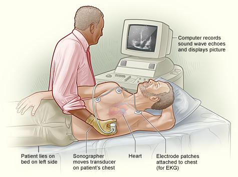
An echocardiogram is a test that uses ultrasound to evaluate your heart muscle and heart valves.

Why Do I Need an Echocardiogram?

Your doctor may perform an echocardiogram to:

  • Assess the overall function of your heart
  • Determine the presence of many types of heart disease
  • Follow the progress of heart valve disease over time
  • Evaluate the effectiveness of medical or surgical treatments

What is a Nuclear stress test?

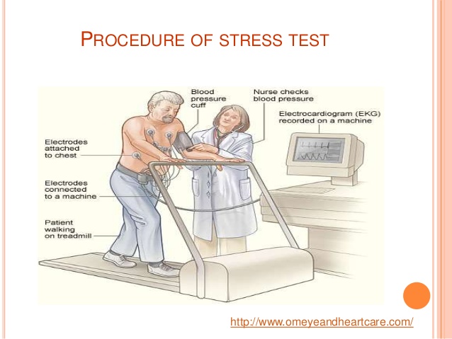
A nuclear stress test helps to determine which parts of the heart are healthy and function normally and which are not. A very small and harmless amount of radioactive substance is injected into the patient. Then the doctor uses a special camera to identify the rays emitted from the substance within the body; this produces clear pictures of the heart tissue on a monitor. These pictures are done both at rest and after exercise. Using this technique, a less than normal amount of the substance will be seen in those areas of the heart that have a decreased blood supply.

What is a Mammogram?

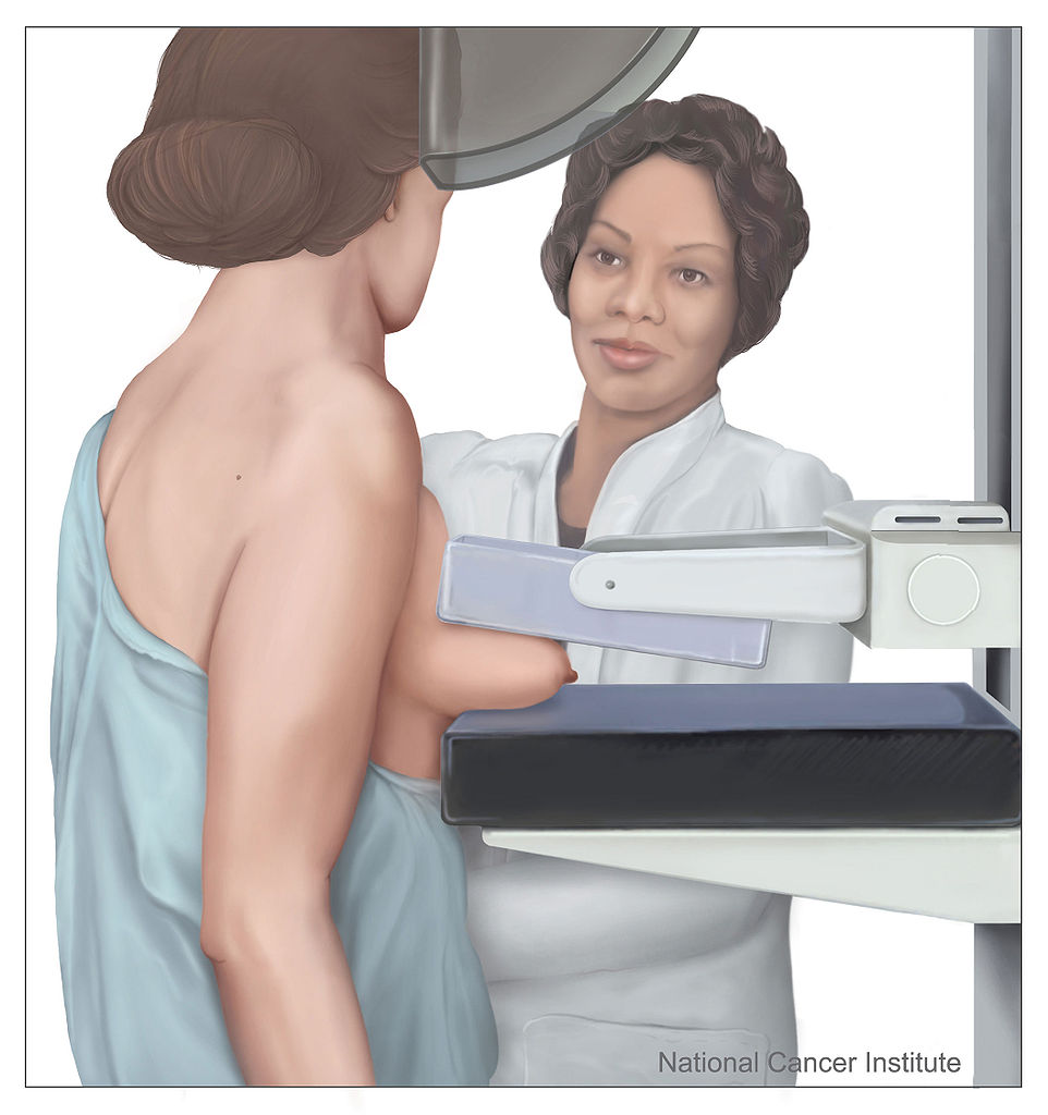
Mammography is a specific type of breast imaging that uses low-dose x-rays to detect cancer early – before women experience symptoms – when it is most treatable.

Tell your doctor about any breast symptoms or problems, prior surgeries, hormone use, whether you have a family or personal history of breast cancer, and if there’s a possibility you are pregnant. If possible, obtain copies of your prior mammograms and make them available to your radiologist on the day of your exam. Leave jewelry at home and wear loose, comfortable clothing. You may be asked to wear a gown. Don’t wear deodorant, talcum powder or lotion under your arms or on your breasts as these may appear on the mammogram and interfere with correct diagnosis.

How should I prepare for the mammogram?

Before scheduling a mammogram, the American Cancer Society (ACS) and other specialty organizations recommend that you discuss any new findings or problems in your breasts with your doctor. In addition, inform your doctor of any prior surgeries, hormone use, and family or personal history of breast cancer.

Do not schedule your mammogram for the week before your menstrual period if your breasts are usually tender during this time. The best time for a mammogram is one week following your period. Always inform your doctor or x-ray technologist if there is any possibility that you are pregnant.

The ACS also recommends you:

  • Do not wear deodorant, talcum powder or lotion under your arms or on your breasts on the day of the exam. These can appear on the mammogram as calcium spots.
  • Describe any breast symptoms or problems to the technologist performing the exam.
  • Obtain your prior mammograms and make them available to the radiologist if they were done at a different location. This is needed for comparison with your current exam and can often be obtained on a CD.
  • Ask when your results will be available; do not assume the results are normal if you do not hear from your doctor or the mammography facility.

What is a Xray?

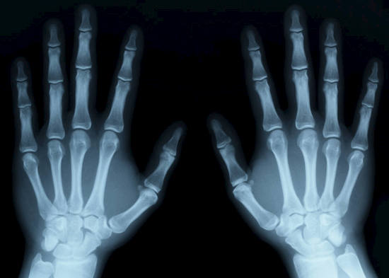
X-rays are a type of radiation called electromagnetic waves. X-ray imaging creates pictures of the inside of your body. The images show the parts of your body in different shades of black and white. This is because different tissues absorb different amounts of radiation. Calcium in bones absorbs x-rays the most, so bones look white. Fat and other soft tissues absorb less, and look gray. Air absorbs the least, so lungs look black.

The most familiar use of x-rays is checking for broken bones, but x-rays are also used in other ways. For example, chest x-rays can spot pneumonia. Mammograms use x-rays to look for breast cancer.

When you have an x-ray, you may wear a lead apron to protect certain parts of your body. The amount of radiation you get from an x-ray is small. For example, a chest x-ray gives out a radiation dose similar to the amount of radiation you’re naturally exposed to from the environment over 10 days.

What is a Hearing Test?

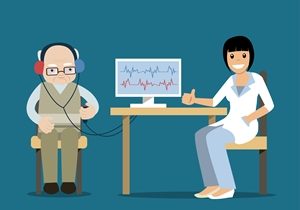
Every now and then, your doctor may ask you to get a hearing test. Don’t assume that something is wrong. Hearing tests are how doctors use to make sure that your ears work well.

With age, hearing loss becomes more likely. About 14% of people ages 45 to 64 have some degree of hearing loss, but that rises to more than 30% among people who are 65 or older. This is why your doctor will want to test your hearing every few years, rather than just once as an adult.

Experts recommend that adults get their hearing tested every 10 years until age 50, and then every 3 years after that.

Why Would I Need a Hearing Test?

Some people may suspect that they have hearing loss. They have trouble hearing people talk to them when they’re in a crowded room, or they’ve been told they raise the volume on the TV way too high.

But not all people know that they have a problem. You may not realize that you have hearing loss, because it’s often a gradual process. That’s why it’s important to have your ears checked when your doctor says you should, even if you think you’re fine.

There are many causes of hearing loss in adults:

  • Being around loud noises often at work
  • Mowing the lawn or using power tools
  • Shooting guns or other weapons
  • Loud music, both live and recorded
  • Too much ear wax
  • Getting hit on the head
  • Having an infection
  • Taking certain drugs
  • Problems with hearing that run in the family

Older adults who don’t do anything to address their hearing loss are more likely to feel left out of social events that they’d normally enjoy, because they can’t hear what’s happening. They might even stop seeing their friends or family as often because they’re embarrassed that they can’t hear well. Isolation makes people more likely to become depressed, unless they get help for their hearing loss.

What is a ultrasound?

Ultrasound imaging uses sound waves to produce pictures of the inside of the body. It is used to help diagnose the causes of pain, swelling and infection in the body’s internal organs and to examine a baby in pregnant women and the brain and hips in infants. It’s also used to help guide biopsies, diagnose heart conditions, and assess damage after a heart attack. Ultrasound is safe, noninvasive, and does not use ionizing radiation.

This procedure requires little to no special preparation. Your doctor will instruct you on how to prepare, including whether you should refrain from eating or drinking beforehand. Leave jewelry at home and wear loose, comfortable clothing. You may be asked to wear a gown.

What are some common uses of the procedure?

Ultrasound examinations can help to diagnose a variety of conditions and to assess organ damage following illness.

Ultrasound is used to help physicians evaluate symptoms such as:

  • pain
  • swelling
  • infection

Ultrasound is a useful way of examining many of the body’s internal organs, including but not limited to the:

  • heart and blood vessels, including the abdominal aorta and its major branches
  • liver
  • gallbladder
  • spleen
  • pancreas
  • kidneys
  • bladder
  • uterus, ovaries, and unborn child (fetus) in pregnant patients
  • eyes
  • thyroid and parathyroid glands
  • scrotum (testicles)
  • brain in infants
  • hips in infants
  • spine in infants

Ultrasound is also used to:

  • guide procedures such as needle biopsies, in which needles are used to sample cells from an abnormal area for laboratory testing.
  • image the breasts and guide biopsy of breast cancer
  • diagnose a variety of heart conditions, including valve problems and congestive heart failure, and to assess damage after a heart attack. Ultrasound of the heart is commonly called an “echocardiogram” or “echo” for short.

Doppler ultrasound images can help the physician to see and evaluate:

  • blockages to blood flow (such as clots)
  • narrowing of vessels
  • tumors and congenital vascular malformations
  • reduced or absent blood flow to various organs
  • greater than normal blood flow to different areas, which is sometimes seen in infections

With knowledge about the speed and volume of blood flow gained from a Doppler ultrasound image, the physician can often determine whether a patient is a good candidate for a procedure like angioplasty.

What is a bone density?

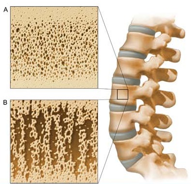
A bone density test is the only test that can diagnose osteoporosis before a broken bone occurs. This test helps to estimate the density of your bones and your chance of breaking a bone. NOF recommends a bone density test of the hip and spine by a central DXA machine to diagnose osteoporosis. DXA stands for dual energy x-ray absorptiometry.

You can find out whether you have osteoporosis or if you should be concerned about your bones by getting a bone density test. Some people also call it a bone mass measurement test. This test uses a machine to measure your bone density. It estimates the amount of bone in your hip, spine and sometimes other bones. Your test result will help your healthcare provider make recommendations to help you protect your bones.

Are you a postmenopausal woman or man age 50 and older? Have you recently broken a bone? If you answered “yes” to both questions, you should talk to your doctor or other healthcare provider about getting a bone density test if you’ve never had one.

What a Bone Density Test Can Do

A bone density test tells you if you have normal bone density, low bone density (osteopenia) or osteoporosis. It is the only test that can diagnose osteoporosis. The lower your bone density, the greater your risk of breaking a bone. A bone density test can help you and your healthcare provider:

  • learn if you have weak bones or osteoporosis before you break a bone
  • predict your chance of breaking a bone in the future
  • see if your bone density is improving, getting worse or staying the same
  • find out how well an osteoporosis medicine is working
  • let you know if you have osteoporosis after you break a bone

What are vaccines?

Vaccines play an important role in keeping us healthy. They protect us from serious and sometimes deadly diseases — like haemophilus influenzaetype b (Hib) and measles.

It’s normal to have questions about vaccines. Vaccines.gov works with scientists and doctors to answer your questions and provide the information you need to get vaccinated.

Once you have the information you need, make sure that you and your family are up-to-date on your vaccinations — they’re your best shot against serious, preventable illness. Find more answers to common questions about vaccines.